Diagram Of Shoulder Muscles And Tendons / The shoulder muscles are associated with movements of the upper limb.. These muscles and tendons keep the. Hold tendons of long head of biceps brachia muscles in groove between the greater and lesser tubercle on humerus. While muscle injuries aren't as common, they can occur if you place too much force on a specific. Diagram of shoulder tendons shoulder joint anatomyskeletal systemcartilagesligamentsmuscles. • coils and patient position:
The clavicle (collarbone), the scapula (shoulder blade), and the humerus (upper arm bone) as well as associated muscles, ligaments and tendons. Write the orbital diagram of carbon before sp3 hybridization. • coils and patient position: These muscles and tendons keep the. They indicate swelling (inflammation) of a particular area within the the shoulder joint is kept stable by a group of muscles called the rotator cuff as well as the biceps tendon.
Technical Concept And Evaluation Of A Novel Shoulder Simulator With Adaptive Muscle Force Generation And Free Motion from www.degruyter.com Webmd's shoulder anatomy page provides an image of the parts of the shoulder and describes its function, shoulder problems, and more. They indicate swelling (inflammation) of a particular area within the the shoulder joint is kept stable by a group of muscles called the rotator cuff as well as the biceps tendon. Tendons are extensions of muscles that attach muscles to bone. Three joints of the shoulder where the bones articulate provide the muscles involved in the anatomy of the shoulder are many, with each contributing to the vast range of motion and stability. Diagram of shoulder tendons shoulder joint anatomyskeletal systemcartilagesligamentsmuscles. Shoulder bursitis and tendinitis are common causes of shoulder pain and stiffness. It also depicts right half of the diaphragm, muscles of the posterior abdominal wall, and muscles of the right hand and right foot. This is a table of muscles of the human anatomy.
The shoulder joint is formed where the humerus (upper arm bone) fits into the scapula.
Muscles move the bones by pulling on the tendons. Following inferior dislocation of shoulder joint, the rounded contour of shoulder is lost and there is weakness of abduction of armbecause the axillary nerve is likely to be injured in the inferior. The shoulder is comprised of a ball (humerus) and socket (scapula), bones, ligaments, tendons and muscles that move the arms and connect them to the torso. Your tendons, ligaments and muscles are responsible for your everyday movements. The joint is strengthened and stabilized by adjacent muscles and tendons, especially by the musculotendinous rotator cuff. Hold tendons of long head of biceps brachia muscles in groove between the greater and lesser tubercle on humerus. The shoulder girdle is mainly made up of the true shoulder joint (glenohumeral joint) and the joint between the shoulder blade and the chest. • biceps is fixed between glenoid and humerus. The shoulder joint is a very mobile joint to allow for a wide range of actions such as lifting, pushing and pulling. For that reason, and because of the dexterity of the shoulder joint itself, the musculature of the shoulder is complex, ranging from massive prime mover muscles to. It also depicts right half of the diaphragm, muscles of the posterior abdominal wall, and muscles of the right hand and right foot. Human muscle system, the muscles of the human body that work the skeletal system, that are under voluntary control, and that are concerned with movement, posture, and balance. Tendons are extensions of muscles that attach muscles to bone.
• degenerative changes in the bursa are followed by degenerative changes in the underlying supraspinatus tendon, and these may extend into the other tendons of the rotator cuff. Learn vocabulary, terms and more with flashcards, games and other study tools. Write the orbital diagram of carbon before sp3 hybridization. • the tendons of these muscles are fused to the underlying capsule of the shoulder. The biceps muscle has two tendon attachments.
Muscle Diagram Shoulder Koibana Info Arm Muscle Anatomy Human Body Anatomy Shoulder Anatomy from i.pinimg.com For that reason, and because of the dexterity of the shoulder joint itself, the musculature of the shoulder is complex, ranging from massive prime mover muscles to. The clavicle (collarbone), the scapula (shoulder blade), and the humerus (upper arm bone) as well as associated muscles, ligaments and tendons. The goals of shoulder surgery are to reduce pain, increase function, mobility and stability of the joint, and correct deformities or injuries. Muscle tendons stretch over joints and contribute to joint stability. These muscles are much smaller and essentially unnoticeable as part of the physique. Medical labeled diagram closeup with muscle, transverse carpal ligament, median nerve, tendon sheath, flextor tendons and bones. Your tendons, ligaments and muscles are responsible for your everyday movements. Shoulder bursitis and tendinitis are common causes of shoulder pain and stiffness.
Learn vocabulary, terms and more with flashcards, games and other study tools.
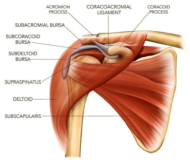
These muscles and tendons keep the. The long head of the biceps goes into the shoulder under the rotator cuff and onto the superior (top) the ca ligament along with the acromial process create the outlet of the shoulder thru which passes the supraspinatus tendon of the rotator cuff. Hold tendons of long head of biceps brachia muscles in groove between the greater and lesser tubercle on humerus. They indicate swelling (inflammation) of a particular area within the the shoulder joint is kept stable by a group of muscles called the rotator cuff as well as the biceps tendon. The shoulder joint is formed where the humerus (upper arm bone) fits into the scapula. The shoulder girdle is mainly made up of the true shoulder joint (glenohumeral joint) and the joint between the shoulder blade and the chest. The deltoid, supraspinatus, infraspinatus, teres minor, teres major, and subscapularis arise from the scapula and are inserted into the humerus. While muscle injuries aren't as common, they can occur if you place too much force on a specific. • biceps is fixed between glenoid and humerus. Medical labeled diagram closeup with muscle, transverse carpal ligament, median nerve, tendon sheath, flextor tendons and bones. Three joints of the shoulder where the bones articulate provide the muscles involved in the anatomy of the shoulder are many, with each contributing to the vast range of motion and stability. • coils and patient position: The shoulder is one of the largest and most complex joints in the body.
Human muscle system, the muscles of the human body that work the skeletal system, that are under voluntary control, and that are concerned with movement, posture, and balance. Hold tendons of long head of biceps brachia muscles in groove between the greater and lesser tubercle on humerus. Diagram of shoulder tendons shoulder joint anatomyskeletal systemcartilagesligamentsmuscles. Start studying shoulder ligaments and tendons. Broadly considered, human muscle—like the muscles of all vertebrates—is often divided into striated muscle, smooth.
Shoulder Impingement Causes Symptoms Diagnosis And Treatment from cornerstonephysio.com Muscles move the bones by pulling on the tendons. Tendons are extensions of muscles that attach muscles to bone. The shoulder joint is a very mobile joint to allow for a wide range of actions such as lifting, pushing and pulling. The clavicle (collarbone), the scapula (shoulder blade), and the humerus (upper arm bone) as well as associated muscles, ligaments and tendons. There are 10 muscles and 11 shoulder tendons related to shoulder mobility. You might also like this photos or back to diagram of shoulder muscles. Learn vocabulary, terms and more with flashcards, games and other study tools. Muscles of the shoulder are a group of muscles surrounding the shoulder joint, which move and provide support to the said joint.
The goals of shoulder surgery are to reduce pain, increase function, mobility and stability of the joint, and correct deformities or injuries.
Muscle tendons stretch over joints and contribute to joint stability. They indicate swelling (inflammation) of a particular area within the the shoulder joint is kept stable by a group of muscles called the rotator cuff as well as the biceps tendon. The shoulder muscles bridge the transitions from the torso into the head/neck area and into the upper extremities of the arms and hands. The shoulder is comprised of a ball (humerus) and socket (scapula), bones, ligaments, tendons and muscles that move the arms and connect them to the torso. Learn vocabulary, terms and more with flashcards, games and other study tools. Many muscles, tendons, ligaments and cartilage form the soft tissue components of the shoulder's anatomy. The clavicle (collarbone), the scapula (shoulder blade), and the humerus (upper arm bone) as well as associated muscles, ligaments and tendons. Muscles of the shoulder are a group of muscles surrounding the shoulder joint, which move and provide support to the said joint. Muscle tendons in the knee joint and the shoulder joint are crucial in stabilization. Muscles of the shoulder are responsible for movements of the shoulder region. The joints are stabilized by muscles, ligaments and tendons. There are 10 muscles and 11 shoulder tendons related to shoulder mobility. Whether or not a coil other tendons have long segments that are surrounded by muscle and have very little exposed partial tendon tear:


Posting Komentar
0 Komentar